Back Of Neck Anatomy - Labeled Anatomy Chart Of Neck And Back Photograph By Hank Grebe. The larynx is located where the pharynx, the back of the mouth and nasal cavity, divides into the trachea (the tube that carries air to the lungs) and the esophagus (the tube that carries food to. Muscle head anatomy vocal organ diagram female neck anatomy neck wireframe head neck human anatomy head artery anatomy face pharynx vector neck degree head anatomy 3d. These muscles also flex the head, and from their obliquity, rotate it, so as to turn the face to one or the other side. The muscles of the back muscles make up a large part of the anatomy (structure) of the back. Anatomy of back of human neck, anatomy of the back and neck, anatomy of the back of the neck, anatomy of the back of the neck muscles, anatomy of the back of your.
Causes of neck pain and how to manage the pain in basic terms, the neck (cervical spine) joins the shoulders and chest to the head. The back anatomy includes the latissimus dorsi, trapezius, erector spinae, rhomboid, and the teres major. Neck anatomy nerves picture there are 8 spinal nerves that originate from the cervical spine. Neck anatomy explained the neck begins at the base of the skull and connects to the thoracic spine (the upper back). The external jugular veins descend on either side of the neck, passing over the sternomastoid muscles and beneath the platysma.
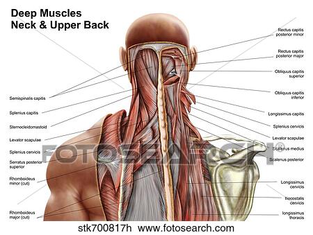
Human Anatomy Showing Deep Muscles In The Neck And Upper Back Drawing Stk700817h Fotosearch from fscomps.fotosearch.com Neck muscles can be strained from poor posture — whether it's leaning over your computer or hunching over your workbench. The external carotid artery supplies the areas of the head and neck external to the cranium. Neck anatomy nerves picture there are 8 spinal nerves that originate from the cervical spine. Osteoarthritis also is a common cause of neck pain. Jugularis posterior) begins in the occipital region and returns the blood from the skin and superficial muscles in the upper and back part of the neck, lying between the splenius and trapezius. Contains glands ( thyroid , parathyroid, and thymus ), the larynx , pharynx and trachea. The content of the neck is grouped into 4 neck spaces, called the compartments. The neck muscles, including the sternocleidomastoid and the trapezius, are responsible for the gross motor movement in the muscular system of the head and neck.
The top of the cervical spine connects to the skull, and the bottom connects to the upper back at about shoulder level.
Neck muscles can be strained from poor posture — whether it's leaning over your computer or hunching over your workbench. Causes of neck pain and how to manage the pain in basic terms, the neck (cervical spine) joins the shoulders and chest to the head. The skull is a strong, bony capsule that rests on the neck and encloses the brain. The muscles of the back muscles make up a large part of the anatomy (structure) of the back. The longus capitis and rectus capitis anterior are the direct antagonists of the muscles at the back of the neck, serving to restore the head to its natural position after it has been drawn backward. The neck muscles, including the sternocleidomastoid and the trapezius, are responsible for the gross motor movement in the muscular system of the head and neck. The external carotid artery supplies the areas of the head and neck external to the cranium. The back of the neck is mostly comprised of muscles, as well as the spine. After arising from the common carotid artery, it travels up the neck, passing posteriorly to the mandibular neck and anteriorly to the lobule of the ear. Neck anatomy nerves picture there are 8 spinal nerves that originate from the cervical spine. Neck anatomy explained the neck begins at the base of the skull and connects to the thoracic spine (the upper back). The occipital bone surrounds a large opening known as the foramen magnum. These muscles also flex the head, and from their obliquity, rotate it, so as to turn the face to one or the other side.
The neurocranium (cranial vault) and the viscerocranium (facial skeleton). Neck muscles can be strained from poor posture — whether it's leaning over your computer or hunching over your workbench. The cervical spine, your neck, is a complex structure making up the first region of the spinal column starting immediately below the skull and ending at the first thoracic vertebra. An area called the occiput. Jugularis posterior) begins in the occipital region and returns the blood from the skin and superficial muscles in the upper and back part of the neck, lying between the splenius and trapezius.
Human Torso Model Life Size Torso Model Anatomical Teaching Torso Unisex Torso Open Back Torso 18 Part Torso Model from www.a3bs.com The muscles of the neck are present in four main groups. Contains cervical vertebrae and postural muscles. Muscle head anatomy vocal organ diagram female neck anatomy neck wireframe head neck human anatomy head artery anatomy face pharynx vector neck degree head anatomy 3d. After arising from the common carotid artery, it travels up the neck, passing posteriorly to the mandibular neck and anteriorly to the lobule of the ear. The nerves of the head and neck include the most vital and important organs of the nervous system — the brain and spinal cord — as well as the organs of the special senses. The majority of these nerves control the functions of the upper extremities and allow you to feel your arms, shoulder, and back of your head. The neck is essentially a passageway for air, food, liquids, blood, and more to travel between the head and the rest of the body, through structures such as blood vessels, nerves, and lymph nodes, as well as the larynx, trachea, and esophagus. In the neck are the thyroid and parathyroid glands, that secrete hormones that control metabolism and blood calcium levels.
They empty into the right and left subclavian veins in the base of the neck.
Cervical spine anatomy video the cervical spine has 7 stacked bones called vertebrae, labeled c1 through c7. The occipital bone surrounds a large opening known as the foramen magnum. Osteoarthritis also is a common cause of neck pain. After arising from the common carotid artery, it travels up the neck, passing posteriorly to the mandibular neck and anteriorly to the lobule of the ear. The external carotid artery supplies the areas of the head and neck external to the cranium. In addition, in this region we also find the major cranial and spinal nerves that connect the central nervous system to the organs, skin, and muscles of the head and neck. The internal jugular veins form the major venous drainage of the head and neck and are deep veins that parallel the common carotid artery. Top head neck anatomy flashcards ranked by quality. In the neck are the thyroid and parathyroid glands, that secrete hormones that control metabolism and blood calcium levels. Occipital neuralgia is caused due to irritation or injury to the occipital nerve. The occipital bone is the only bone in your head that connects with your cervical spine (neck). Think of it like a jigsaw puzzle, all the pieces fit in together and are required to get the full picture as to how it works. They start at the top of the neck and go down to the tailbone.
Search for anatomy back neck. Osteoarthritis also is a common cause of neck pain. The occipital bone is a bone that covers the back of your head; Occipital neuralgia is caused due to irritation or injury to the occipital nerve. The longus capitis and rectus capitis anterior are the direct antagonists of the muscles at the back of the neck, serving to restore the head to its natural position after it has been drawn backward.
Vivian Grisogono About The Back And Neck from www.viviangrisogono.com The posterior external jugular vein (v. The internal jugular veins form the major venous drainage of the head and neck and are deep veins that parallel the common carotid artery. Muscle head anatomy vocal organ diagram female neck anatomy neck wireframe head neck human anatomy head artery anatomy face pharynx vector neck degree head anatomy 3d. It is made up of bones, discs, muscles, ligaments, nerves and tendons. Occipital neuralgia is caused due to irritation or injury to the occipital nerve. The cervical spine supports the weight and movement of your head and protects the nerves exiting your brain. The external jugular veins descend on either side of the neck, passing over the sternomastoid muscles and beneath the platysma. These muscles give the sides of the neck their.
It consists of two major parts:
They empty into the right and left subclavian veins in the base of the neck. In this video, i walk you through a basic approach to drawing the neck and upper back muscles. Contains cervical vertebrae and postural muscles. The skull is a strong, bony capsule that rests on the neck and encloses the brain. Think of it like a jigsaw puzzle, all the pieces fit in together and are required to get the full picture as to how it works. The muscles of the back muscles make up a large part of the anatomy (structure) of the back. Contains glands ( thyroid , parathyroid, and thymus ), the larynx , pharynx and trachea. The occipital bone surrounds a large opening known as the foramen magnum. The top of the cervical spine connects to the skull, and the bottom connects to the upper back at about shoulder level. The external jugular veins descend on either side of the neck, passing over the sternomastoid muscles and beneath the platysma. In addition, in this region we also find the major cranial and spinal nerves that connect the central nervous system to the organs, skin, and muscles of the head and neck. Rarely, neck pain can be a symptom of a more serious problem. Muscle head anatomy vocal organ diagram female neck anatomy neck wireframe head neck human anatomy head artery anatomy face pharynx vector neck degree head anatomy 3d.